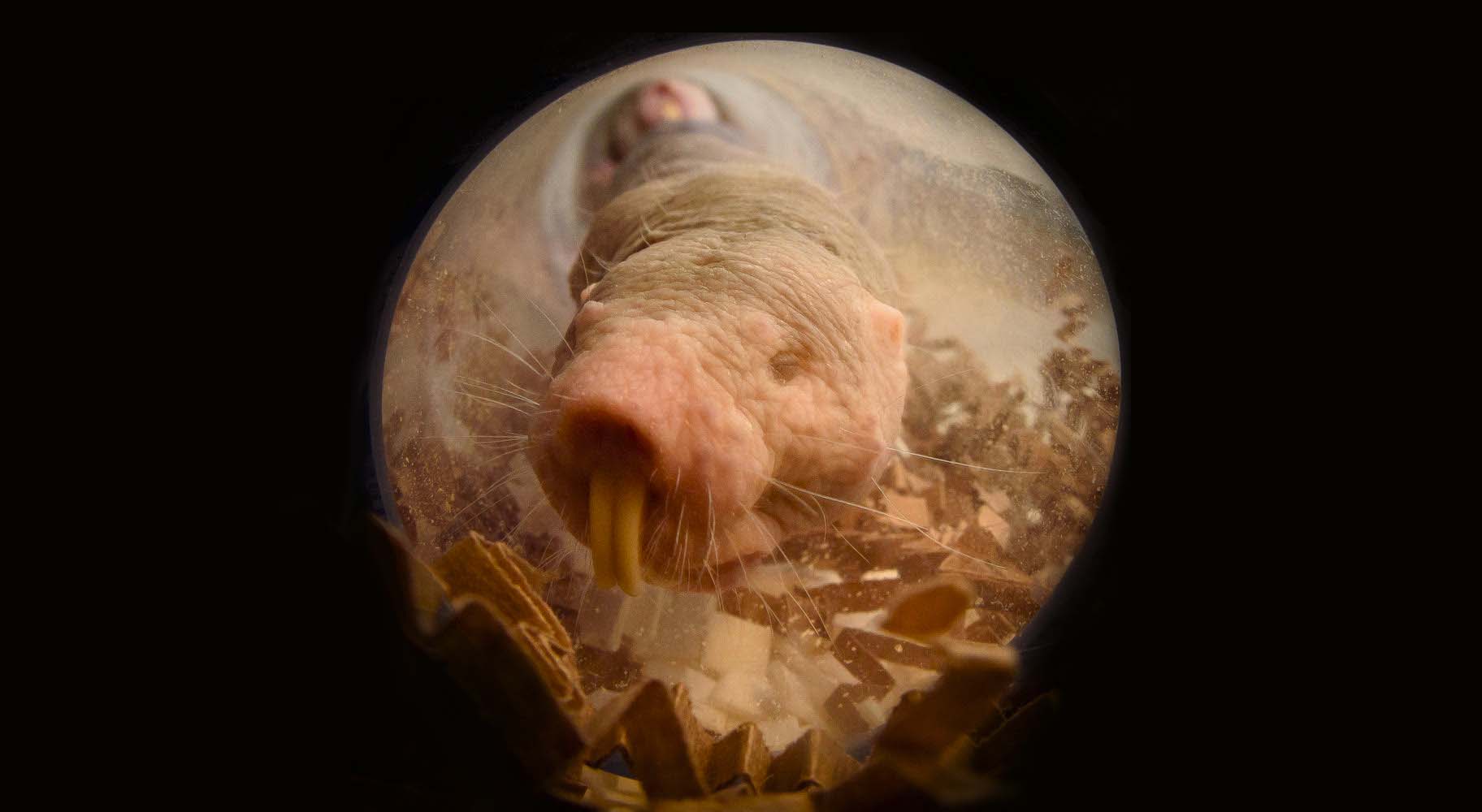
NAKED AND UNAFRAID TO THERMOREGULATE
For University of Ottawa researcher Matthew Pamenter, naked mole rats were the perfect candidate to explore how animals have evolved to live in very low levels of oxygen (hypoxic environments). His research, titled “Naked mole-rat brown fat thermogenesis is diminished during hypoxia through a rapid decrease in UCP1,” was published in Nature Communications.
This week, Pamenter interviewed with the Fulcrum to explain this mechanism that enables mole rats to decrease their body temperature in hypoxia.
How do naked mole rats create hypoxic environments?
According to the researcher, mole rats are a eusocial species, meaning that they live together in a colony, and at one time there could be hundreds of them living together. Mole rats tend to burrow underground and create a vast network of tunnels.
When a lot of animals live in a small and confined space, “they’re first breathing and thus use up the oxygen. It’s thought that they create a hypoxic environment for themselves within their nest chamber. As they leave their nests through the tunnel systems they enter a normoxic environment where there is more oxygen,” said Pamenter.
As a result, mole rats live in and out of hypoxic environments and experience intermittent hypoxic stress.
How do most hypoxia-tolerant animals survive?
Pamenter explained that “we [humans] use oxygen at the mitochondrial level to generate aerobic energy, so to make ATP (energy) that our cells need. When we don’t have enough oxygen our cells can’t produce energy.”
This means that we can either increase the anaerobic metabolic pathways which is when we produce ATP without oxygen or we can turn down our metabolic demand. For naked mole rats, thermoregulation is one of the most energetically expensive processes. Since they lack fur, it’s difficult for them to maintain their body temperature.
What Pamenter found in previous studies looking at what mole rats do with their body temperature in hypoxic conditions was that they do in fact decrease their body temperature in hypoxia. In his most recent study, he examines the mechanism that explains the phenomenon that allows mole rats to reduce their body temperature in hypoxic environments.
Understanding the mechanism
According to Pamenter, small rodents have a fat tissue called brown fat, which contains specialized mitochondria that contain a protein called uncoupling protein one (UCP1). During the process of oxidative phosphorylation (a metabolic pathway), the electron transport chain and mitochondria pump protons across the mitochondrial membrane. In doing so, they build up a proton motive force, stored as potential energy.
He continued, “think of this as like a sump pump, which is pumping protons to an elevated level (creating a proton gradient) and then the ATP synthase uses that to phosphorylate, ADP to ATP. This is the mechanism that our cells use to make ATP.”
Now consider the uncoupling protein, which “essentially puts a bunch of holes in that membrane that lets the protons just flow back across it without actually generating ATP. By uncoupling the hydrogen gradient from ATP production, you get this futile cycle where the mitochondria are constantly pumping protons, and then they’re leaking back across. As a result, this generates heat and so these fat cells generate heat, and this is called non-shivering thermogenesis.”
When small rodents need to produce heat they are able to turn these proteins (UPC 1 in brown fat) on, and by doing so they generate lots of heat in the regions containing this specialized fat. This process is called non-shivering thermogenesis due to the fact that the rodents can produce heat without making the muscle shiver.
Contrast this process with humans: when we are cold our bodies will shiver and our muscles will twitch, generating heat.
What were the findings of this study?
Most interestingly, according to the researcher, “mole rats do express this protein (UCP1) in their brown fat, but when they’re in hypoxia this protein is downregulated very rapidly from their brown fat membranes. This correlates with a decrease in heat production in their interscapular region (between the shoulder blades).”
Now consider mice, who take three to four days for those proteins to be cycled and removed from the membrane.
In addition to describing the mechanism which explains how naked mole rats can save energy to survive in hypoxia, Pamenter and his team studied several different but related species of mole rats that lived in South Africa.
What they found was that “in other social species [the ones found in South Africa] UCP1 also decreased with hypoxia quite quickly. Whereas in a solitary species it didn’t. This suggests that the ability to turn off UCP1 so fast and so much faster than mice may have evolved preferentially in the social species.”
What’s next for Pamenter?
In the future, Pamenter is looking forward to examining pathways in the brain to see if there are changes in the mitochondrial networks and how this impacts mitochondrial energetics, which may support hypoxia tolerance and improved energy production in brain tissue in hypoxia.